


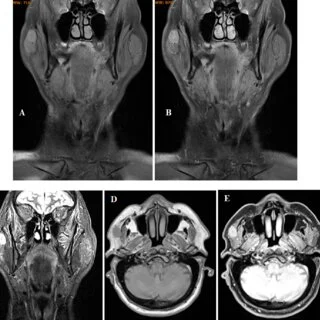
Body imaging uses CT scans, MRI, ultrasound, and X-rays to examine internal organs, soft tissues, bones, and blood vessels. These imaging techniques help physicians identify abnormalities associated with infections, including inflammation, abscesses, and fluid accumulation. Because infections sometimes produce nonspecific symptoms, physicians use imaging findings alongside laboratory tests and medical history to evaluate possible causes. Here is more information about body imaging in evaluating infectious diseases:
Detecting Abdominal Infections
Abdominal and pelvic infections may affect internal organs and surrounding tissues, sometimes producing symptoms that overlap with other conditions. CT scans and ultrasound provide images of abdominal structures, while radiologists examine findings associated with inflammation, abscesses, and fluid accumulation. Because physical examinations cannot directly visualize internal organs, imaging provides additional information when investigating possible infections.
Physicians use abdominal imaging to evaluate the liver, kidneys, intestines, and other structures for signs of infection. CT scans help identify deeper fluid collections, while ultrasound examines selected organs and surrounding tissues without ionizing radiation. When infections involve pelvic structures, imaging findings help physicians determine the affected areas and guide further evaluation. These results also support decisions about medication, drainage procedures, or additional medical care.
Evaluating Musculoskeletal Infections
Musculoskeletal infections may affect bones, joints, and surrounding soft tissues, causing symptoms such as pain, swelling, or limited movement. MRI provides detailed images of these structures, while physicians use the findings to investigate conditions such as osteomyelitis and septic arthritis. Because MRI does not use ionizing radiation, it offers another imaging option when evaluating suspected infections. X-rays also help assess bone abnormalities, although additional imaging may be necessary to identify early infections or soft tissue involvement.
Common imaging applications include:
- Detecting bone infections
- Evaluating joint inflammation
- Identifying soft tissue abscesses
- Examining surrounding tissue damage
- Assessing structural bone changes
Assessing Cardiovascular Infections
Cardiovascular infections may affect the heart, blood vessels, and surrounding tissues, sometimes causing inflammation or structural abnormalities. CT and MRI provide detailed images of cardiovascular structures, while physicians use these findings to investigate suspected vascular infections. Because conditions such as endocarditis involve heart valves, echocardiography often serves as the primary imaging method for evaluation. Additional imaging may help identify complications, examine surrounding tissues, and distinguish infections from other cardiovascular conditions.
Comparing Imaging Exam Types
Body imaging uses different technologies to evaluate infections affecting organs, bones, joints, and surrounding tissues. Physicians select imaging procedures based on symptoms and suspected infection locations, while each technique provides different diagnostic information. Common imaging examinations include:
- CT scans: CT imaging produces cross-sectional images that help identify abdominal abscesses, inflammation, and abnormal fluid collections.
- MRI: MRI uses magnetic fields and radio waves to examine soft tissues, joints, and bones without ionizing radiation.
- Ultrasound: Ultrasound uses sound waves to visualize internal organs, blood vessels, and fluid collections, particularly within the abdomen and pelvis.
- X-rays: X-ray imaging provides images of bones and selected internal structures to evaluate fractures, infections, and other abnormalities.
Because imaging techniques differ in their capabilities, physicians match examinations to the patient’s medical history and clinical findings. CT scans may help investigate suspected appendicitis, while MRI often provides additional information when evaluating possible bone or joint infections.
Supporting Early Diagnosis
Body imaging helps physicians identify abnormalities associated with infections, including abscesses, inflammation, and fluid accumulation. CT scans, MRI, and ultrasound provide detailed information about affected tissues, while imaging findings support diagnostic evaluations. Because infections may change over time, physicians sometimes compare follow-up scans with previous imaging results. These comparisons help assess changes in infection-related abnormalities and guide decisions about further testing or treatment adjustments.
Ask About Body Imaging
Body imaging provides detailed information about internal structures and abnormalities associated with infections. CT scans, MRI, ultrasound, and X-rays help physicians evaluate affected tissues, while imaging findings support diagnostic and treatment decisions. Because symptoms vary between infections, healthcare providers recommend imaging examinations based on medical history and clinical findings. Schedule a body imaging appointment today to complete your recommended evaluation and discuss the results with your healthcare provider.